ColorNephroNet: Kidney Tumour Malignancy Prediction Using Medical Image Colourisation
A two-stage CNN pipeline that colourises grayscale kidney CT scans into pseudo-RGB images to improve malignancy prediction by leveraging features learned from colour image datasets.
Motivation
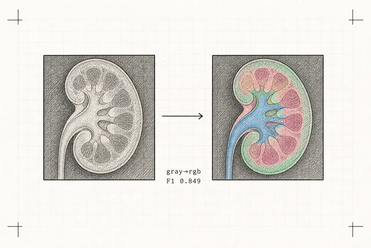
Many nephrectomies (kidney removals) are performed for tumours later found to be benign. We set out to build a system that could support radiological diagnosis by predicting the malignancy of a renal tumour directly from CT images. Conventional CNN models take grayscale CT images as input; we asked whether colourising the medical images into pseudo-RGB could improve classifier performance by allowing the model to leverage features learned from colour image datasets.
Data and Preprocessing
We used CT scans of kidneys with tumours, each with a known benign or malignant outcome confirmed postoperatively. The kidney and tumour region was cropped from each image, intensity values were standardised from the Hounsfield scale to [0,1], images were resized to a fixed resolution, and random flips, rotations, and scaling were applied for augmentation — all with no patient overlap between train, validation, and test splits.
Method
The pipeline runs in two stages. First, an encoder–decoder CNN converts a single-channel CT crop into a three-channel colourised image using an L2 reconstruction loss, introducing richer texture and contrast cues that enable transfer from pre-trained RGB CNNs. Second, a CNN classifier (initialised from ImageNet) predicts a binary benign/malignant label from the colourised image using binary cross-entropy and early stopping.
Results
ColorNephroNet achieved an F1-score of 0.849 versus 0.831 for the grayscale baseline — an improvement of roughly 1.8 percentage points, alongside gains in accuracy (0.840 vs 0.823), precision, and recall. Models were implemented in PyTorch with the Adam optimizer, a 1e-4 learning rate, batch size 32, and weight decay 1e-5.
Keep exploring
UMIE Datasets
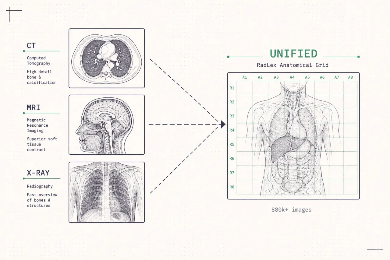
Open-source pipelines that standardize 880k+ images across 20+ medical imaging datasets (CT, MRI, X-ray) into a unified format with RadLex-compliant labels.
View project →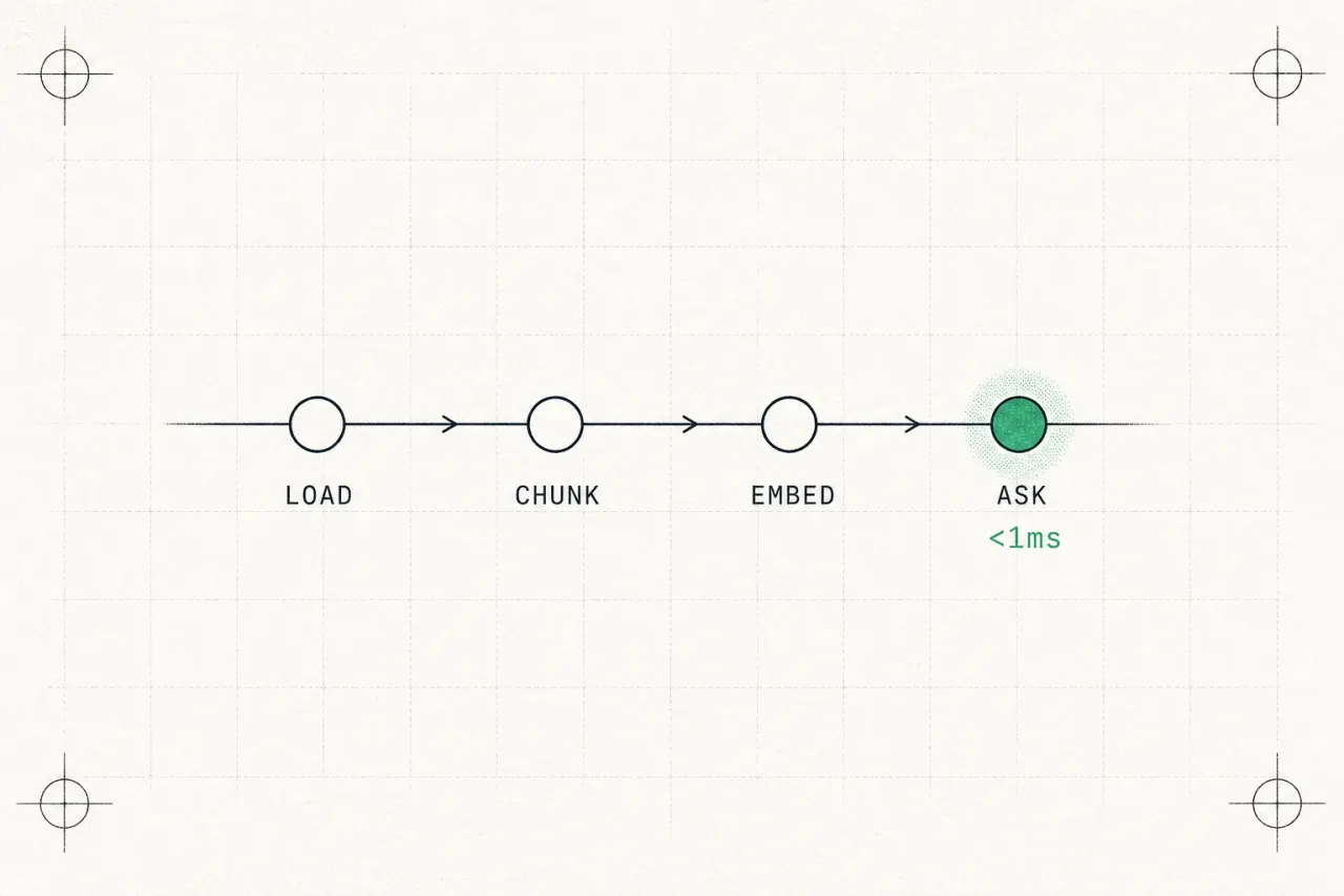
RocketRAG
A speed-focused Retrieval-Augmented Generation framework packaging document ingestion, semantic chunking, vector storage, and LLM inference into a pluggable CLI and FastAPI toolkit.
View project →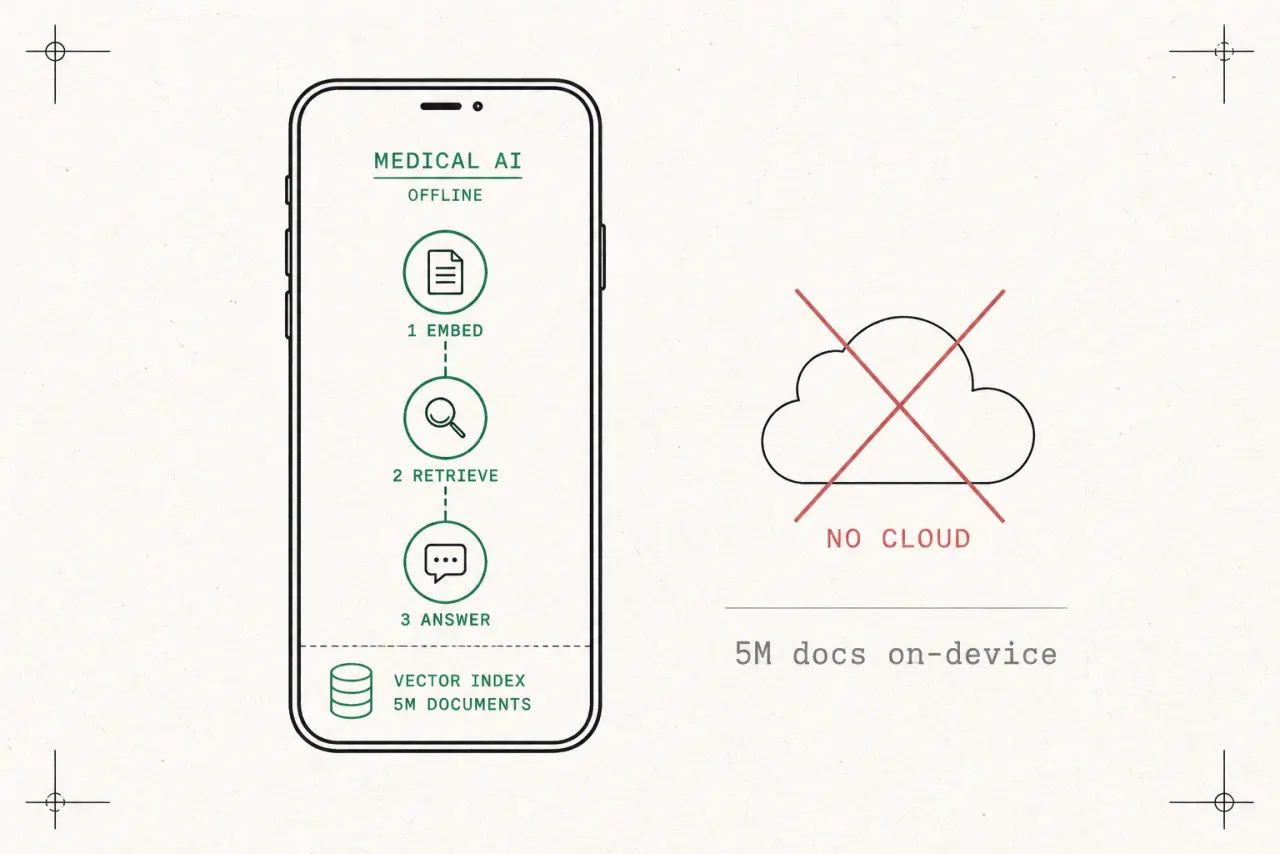
LOMA — Offline Medical AI Assistant
A zero-cloud mobile medical assistant that runs the full pipeline — embeddings, retrieval, and language model responses — entirely on the user's phone.
View project →